
Imaging Services
State-of-the-Art Technology & Equipment
At Touro’s many imaging centers, we diagnose and treat patients using the most advanced technology and methods. Both inpatients and outpatients benefit from the imaging services offered by our experienced and highly trained providers and staff. Read on to learn more about the specific imaging services we offer.
Nuclear medicine
Nuclear medicine is a field of medical study focused on the use of radioactive
substances to diagnose and treat health issues. Radioactive material is
introduced to the body through the mouth or an injection into the patient’s
bloodstream. The patient is monitored by a medical professional using
a special camera and a computer to view and assess the area being examined.
Nuclear medicine detects things that many other imaging procedures cannot,
making it a great way to diagnose diseases in their early stages.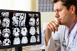
Some of the most common nuclear medicine scans include:
- Renal scans: To examine the kidneys and renal blood flow
- Thyroid scans: To evaluate thyroid function
- Bone scans: To evaluate arthritic changes in joints, to detect bone disease, or to determine the cause of bone pain
- Gallium scans: To diagnose infectious diseases
- Heart scans: To assess blood flow, heart function, and damage to heart muscle after a heart attack
- Brain scans: To assess problems with the brain or blood circulation to the brain
- Breast scans: To locate cancer in the breast
PET scans & CAT scans
PET and CAT scans are two of the most common and most helpful radiology procedures used to identify and diagnose diseases.
A PET (Positron emission tomography) scan is used to examine body tissues or track the progress of treatment while a CAT (computed tomography) scan creates detailed images of the inside of the body. A CAT scan is similar to getting a standard X-ray taken – you may need to ingest iodine dye as a contrast material, but you will not need to ingest any radioactive materials or prepare in any way. Patients who get a PET scan, however, must ingest a small amount of radioactive material so that the doctor can properly locate and study the tissues being examined.
Many patients undergo a PET scan to:
- Diagnose dementias and other neurological conditions
- Locate a surgical site before beginning surgery
- Evaluate the brain after trauma
- Detect whether cancer has spread or to assess the progress of a cancer treatment
PET and CAT scans are often used together so that your doctor can gather more information about cancerous tumors.
MRI
Magnetic resonance imaging (MRI) uses a magnetic field and radio wave energy to examine organs and internal structures. This kind of test tends to provide different information than X-rays and CAT scans. During an MRI, you will be placed in a machine equipped with a strong magnet. A dye may be used as a contrast material. Images are created and saved on a computer so that your doctor can refer back to them. An MRI is most helpful for locating and diagnosing conditions in the brain, organs, glands, blood vessels, and joints.
You may undergo an MRI so that your doctor can look for problems such as:
- Bleeding
- Tumors
- Infection
- Injury
Fluoroscopy
A fluoroscopy is essentially an X-ray “movie” – it studies moving body structures and can evaluate conditions within a specific area of the body.
A fluoroscopy allows your doctor to observe your:
- Skeletal system
- Digestive system
- Urinary system
- Respiratory system
- Reproductive system
The procedure can be done on its own to diagnose diseases or conditions, or it can be used alongside other procedures.
Your doctor may use fluoroscopy to:
- Locate foreign bodies
- Facilitate image-guided anesthetic injections
- Perform intravenous catheter insertion
- Perform percutaneous vertebroplasty (a minimally invasive procedure used to treat compression fractures of the vertebrae of the spine)

